
Figure 2.
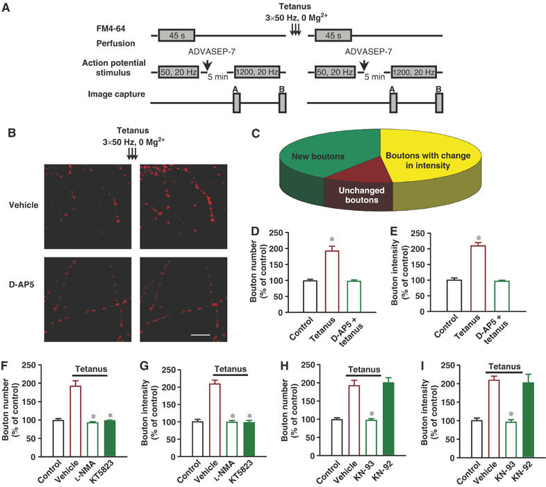
Tetanus in 0 Mg2+ increases functional prS btn number and releasable fluorescence in cultured hippocampal neurons. (A) Experimental protocol for staining, destaining and tetanus application using electrical stimulation. ADVASEP-7 (1 mM), an anionic cyclodextrin complexing agent with higher affinity for the dye than the plasma membrane was introduced in the washing bath solution for enhanced removal of the dye from the external medium to reduce background staining. (B) Examples of activity-dependent FM4-64 staining before and after tetanus. Tetanus in 0 Mg2+ increased prS btn number and releasable fluorescence intensity in pre-existing prS btns. D-AP5 (40 μM) blocked tetanus-induced plasticity. Scale bar 10 μm. (C) A pie chart representing percentage of new btns, btns that underwent changes in fluorescence intensity and unchanged btns, after tetanus (n=5). (D) Percentage increases in number of functional prS btns 30 min after tetanus. D-AP5 (40 μM) completely blocked tetanus-induced increase in recycling btn number (one-way ANOVA followed by LSD). *P<0.001 compared to control group. (E) Percentage increase in releasable fluorescence of pre-existing functional prS btns 30 min after tetanus. D-AP5 (40 μM) blocked tetanus-induced releasable fluorescence increase. *P<0.001 compared to control group. NO synthase inhibitor, L-NMA (50 μM) and cGK inhibitor, KT5823 (2 μM) blocked tetanus-induced increase in prS btn number (F) and releasable fluorescence (G) in pre-existing prS btns. CaMKII inhibitor, KN-93 (5 μM) but not its inactive analog, KN-92 (5 μM) blocked tetanus-induced increase in prS btn number (H) and releasable fluorescence (I) in pre-existing prS btns (one-way ANOVA followed by LSD). *P<0.001 compared to tetanus.