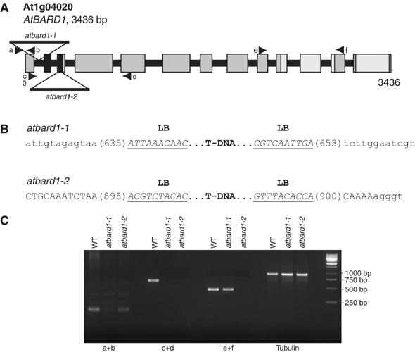
Figure 4.

Schematic structure of the AtBARD1 gene and its T-DNA insertions. (A) The AtBARD1 gene consists of 13 exons. Regions coding for the RING and BRCT domain are indicated in black and light grey, respectively. Two T-DNA insertions were identified. One insertion is located in the first intron, and denominated atbard1-1 whereas the second insertion is located in the third exon, and denominated atbard1-2. (B) An overview of the precise locations of the T-DNA inserts in the AtBARD1 gene. Intron sequences are displayed as lower-case letters, exon sequences as capital letters, and T-DNA border sequences are underlined (LB: left border). (C) Semiquantitative RT–PCR on different regions of the AtBARD1 gene. Primer pairs were used that bind in front of (a+b), across (c+d) and after (e+f) the T-DNA insertions. The β-tubulin gene was taken as control. WT: wild type.
