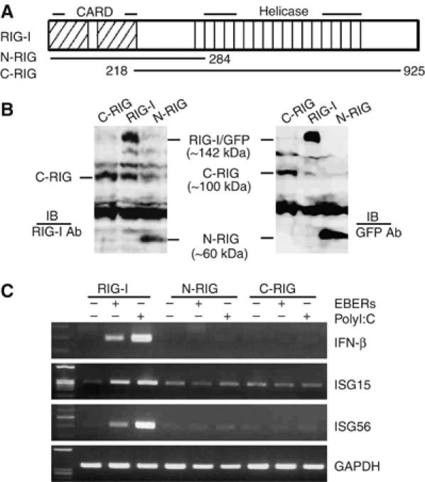
Figure 6.

EBER is recognized by full-length RIG-I. (A) Schematic diagram of mutant plasmids containing the CARD domain (1–284 aa) or the helicase domain (218–925 aa). (B) Immunoblot analysis for detection of mutant RIG-I expressions in EBV-negative Daudi cell clones stably transfected with the deletion plasmids containing GFP-tagged CARD domain (N-RIG) or GFP-tagged helicase domain (C-RIG). Expression of N-RIG and C-RIG was detected by anti-RIG-I antibody (left panel) and also after reprobing with anti-GFP antibody (right panel). (C) Expression of type I IFN and ISGs in EBV-negative Daudi cells stably transfected with the deletion plasmids of RIG-I. The cells (5 × 10⁶ cells each) were transfected with 30 μg of EBERs or polyI:C, and after 6 h, subjected to RT–PCR for detection of IFN-β, ISG15, and ISG56.
