
Figure 4.
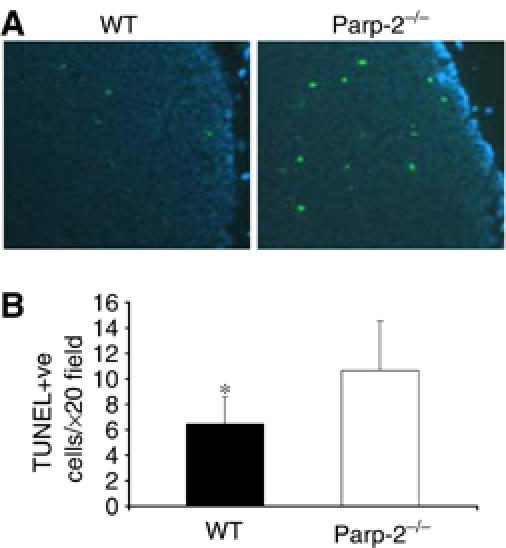
Apoptosis in thymus glands. (A) In situ apoptosis detection in Parp-2−/− and WT mice by the TUNEL assay. Apoptotic cells are labelled in green and cell nuclei are stained with DAPI (blue). (B) Numerical representation of TUNEL-positive (TUNEL +ve) cells. Values represent mean±s.d. for 10 randomly selected cortical fields (n=6 mice per experimental group) (magnification, × 20). *Statistically significant difference (P<0.05).