
Figure 5.
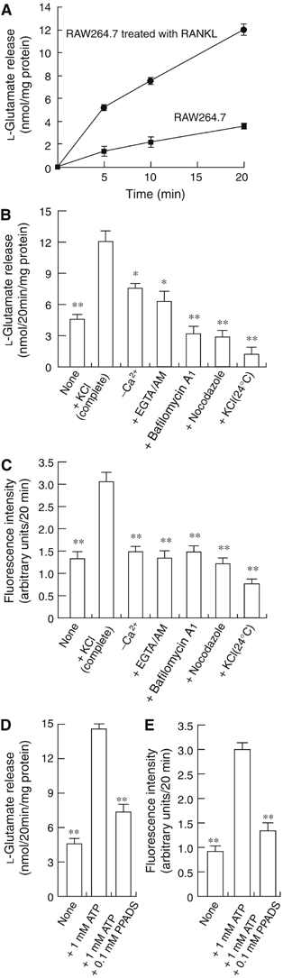
Regulated secretion of L-glutamate and fluorescent bone degradation products from RAW264.7 cells treated with RANKL. (A) RAW264.7 cells treated with RANKL or RAW264.7 cells (2.0 × 10⁵ cells/dish) were stimulated with 50 mM KCl. The L-glutamate released was measured. (B) L-Glutamate secretion after 20 min is shown. In some experiments, cells were treated for 2 h with 1 μM bafilomycin A1, 50 μM EGTA-AM or 10 μM nocodazole, and then stimulated with KCl. (C) Fluorescent bone degradation products in the medium under the conditions in panel B were assessed fluorometrically. The results are means±s.e.m., n=4. Asterisks indicate statistically significant numbers (*P<0.01, **P<0.001). ATP stimulates the secretion of L-glutamate (D) and fluorescent bone degradation products (E) from RAW264.7 cells treated with RANKL. The assay was started by the addition of 1 mM ATP. In some experiments, 0.1 mM PPADS was also included. The results are means±s.e.m., n=4. Asterisks indicate statistically significant numbers (*P<0.01, **P<0.001).