
Figure 8.
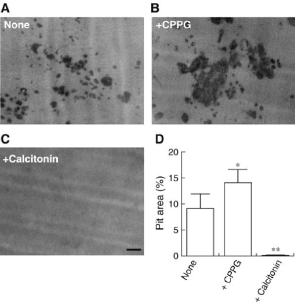
Inhibition of L-glutamate signal stimulated bone resorptive activity. Osteoclasts were cultured on dentine slices in the absence (A) or presence (B) of 100 μM CPPG. In some experiments, 10 nM eel calcitonin was added (C). After 24 h, the resorption pits formed on the dentine slices were stained with Mayer's hematoxylin and quantified using the NIH Image program. The pictures of typical resorption pits formed on the dentine slices under respective conditions are shown. (D) Ratio of resorption area to total area (pit area expressed in %) is shown. *P<0.01, **P<0.001 compared with control. The results are means±s.e.m., n=6. Bar=100 μm.