
Figure 1.
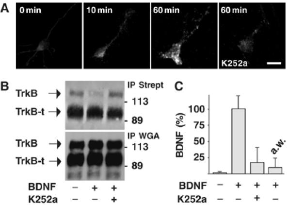
TrkB-mediated BDNF endocytosis in cultured hippocampal neurons. (A) Time course of BDNF endocytosis in untreated neurons (0 min) or neurons exposed to BDNF (10 and 60 min) in the presence or absence of K252a pretreatment, obtained by immunocytochemistry and analyzed by confocal microscopy. Images are representative of 50 neurons analyzed in four independent experiments. Bar, 20 μm. (B) Regulation of plasma membrane expression of TrkB by BDNF. Plasma membrane proteins labeled with activated biotin were isolated by precipitation using agarose-conjugated streptavidin (IP Strept) from lysates of control neurons or neurons treated with BDNF for 60 min. Western blot analysis showed that TrkB, but not TrkB-t, is reduced by BDNF administration; this effect was prevented by pretreating neurons with K252a. Glycoconjugate proteins precipitated by WGA-agarose (IP WGA) showed total amount of TrkB and TrkB-t in whole-cell lysates. (C) Intracellular accumulation of exogenous BDNF measured by ELISA in the same lysates as in panel B. a.w. indicates the amount of BDNF immunoreactivity stripped from the cell surface by acid treatment. Data are expressed as percentage of the means±s.e.m. (n=6).