
Figure 2.
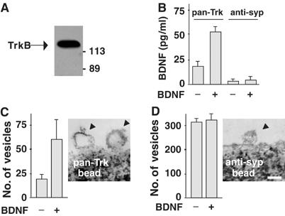
Internalization of BDNF–TrkB complexes in endocytic vesicles. (A) Magnetic beads coated with pan-Trk antibodies (pan-Trk bead) were used to trap Trk-containing vesicles from hippocampal neurons exposed to exogenous BDNF. Western blot analysis using anti-TrkB antibody revealed that purified vesicles express TrkB as a single immunoreactive protein of 135 kDa. (B) ELISA quantification of BDNF in pan-Trk bead and anti-syp bead purifications. Data are means±s.e.m. (n=4). (C) Quantification of vesicles isolated by pan-Trk beads. Vesicle number increased after BDNF exposure for 60 min. Representative image obtained by electron microscopy showing endocytic vesicles attached to a pan-Trk bead. (D) Quantification of vesicles isolated by anti-syp beads. Vesicle number did not change after BDNF exposure for 60 min. Representative image obtained by electron microscopy showing a vesicle with the typical size of a synaptic vesicle attached to an anti-syp bead. Bar, 100 nm. Data in panels C and D are means/100 beads±s.d.