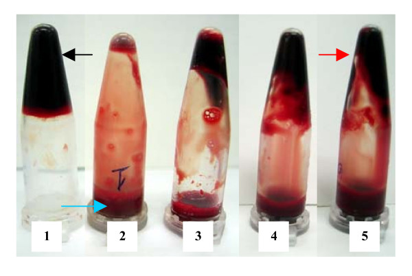
Figure 1.
Clot-lysis of blood samples of normal subjects (positive and negative control). Tube no. 1 is a control clot (negative control) to which water was added. No clot lysis was observed in tube no.1; a black arrow indicates the intact clot. Tube no. 2–5 (positive control) was lysed by four different concentrations of streptokinase with decreasing order. After dissolution of the clots, tubes were inverted and fluid (blue arrow) along with the remnants of clots (red arrow) could be clearly seen

