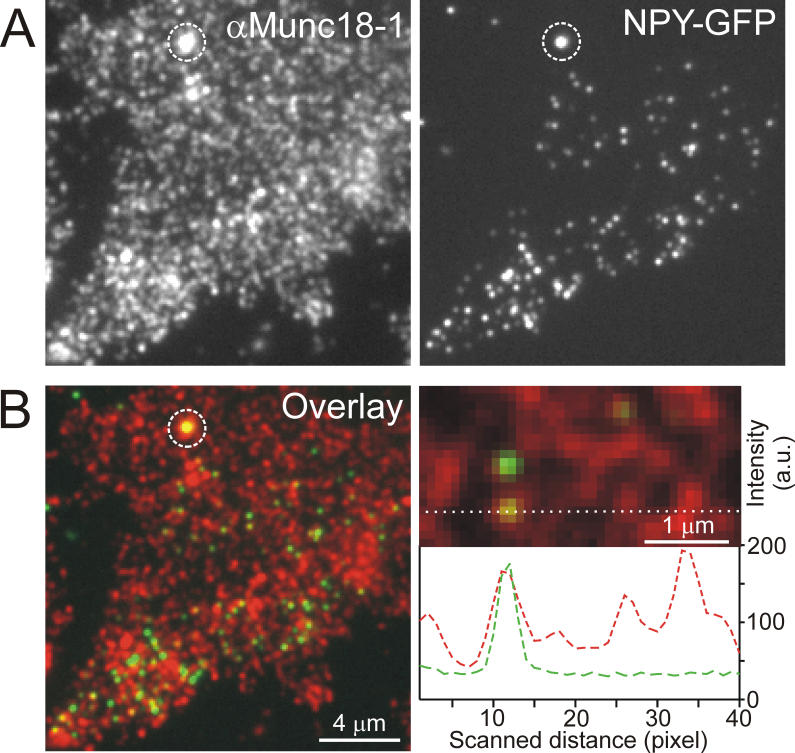
Figure 2. Docking of Secretory Granules to Plasmalemmal Domains Enriched in Munc18–1.
(A) Plasma membrane sheet generated from a PC12 cell expressing the secretory granule marker NPY-GFP. Left, immunostaining for Munc18–1 (red channel); right, plasma membrane–docked, GFP-filled secretory granules (green channel). Circle indicates a fluorescent bead visible in all channels acting as a spatial reference for vertical shifts occurring during filter change.
(B) Left, overlay from images shown in (A); right, magnified view from overlay. Linescans were placed through the centers of individual secretory granules (174 granules from ten membrane sheets were analyzed; for example, see dotted line), and granules were rated to be associated with a Munc18–1–rich domain when both signals had a maximum to within two pixels. Random co-localization was determined on mirrored images and subtracted (for details see Materials and Methods), resulting in 70% specific co-localization of granules with Munc18–1 domains.

