
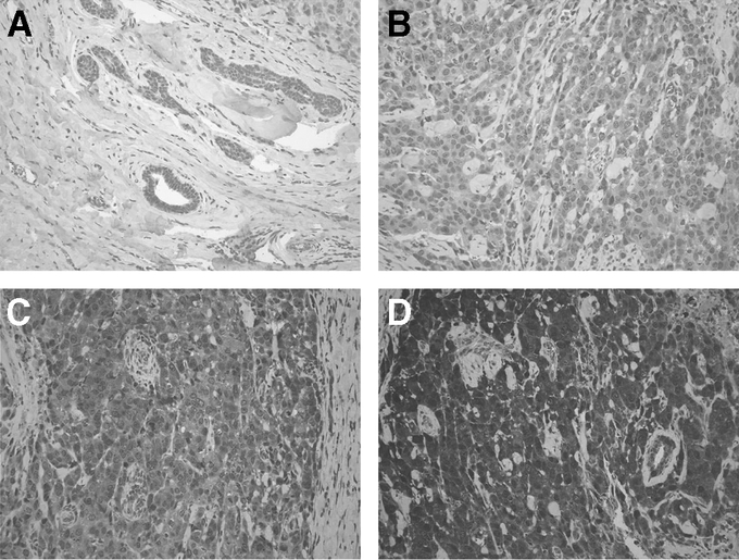
FIGURE 2. Representative slides immunostained for VEGF expression. A, Benign breast tissue, representing grade 0 staining. B–D, Representative panels for 3 breast cancer specimens, demonstrating, respectively, grade I, grade II, and grade III VEGF staining.