
FIGURE 8. Bones of μ-calpain−/− mice are progressively osteopenic.
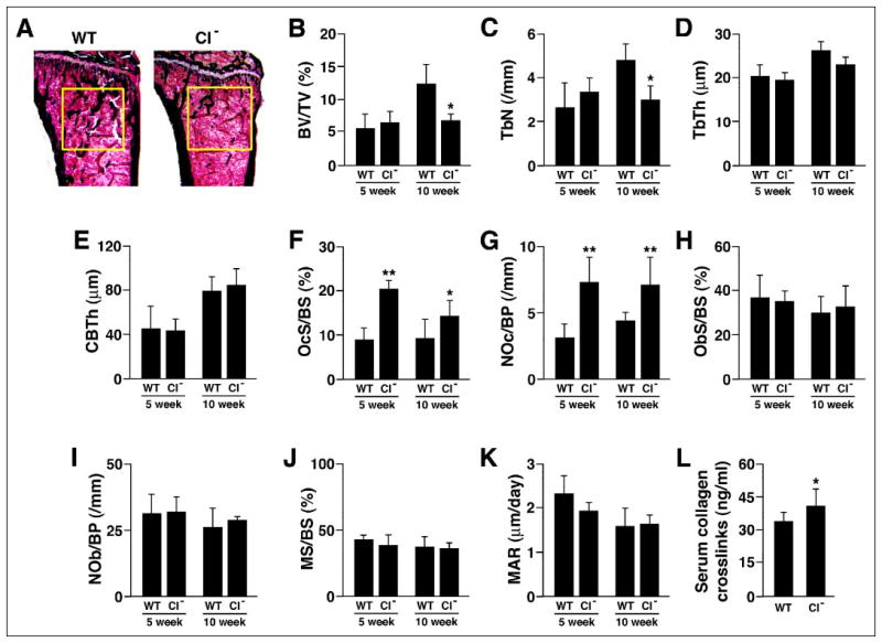
A, Von Kossa staining of tibial proximal metaphyses showing decreased trabeculation of the secondary spongiosa in bones from μ-calpain−/− (CI−) mice. B–E, structural histomorphometry of tibial proximal metaphyses. B, BV/TV, trabecular bone volume; C, TbN, trabecular number; D, TbTh, trabecular thickness; E, CBTh, cortical bone thickness. F–K, cellular and dynamic histomorphometric parameters in tibial proximal metaphyses. F, OcS/BS, osteoclast surface/ bone surface; G, NOc/BP, number of osteoclasts/bone perimeter; H, ObS/BS, osteoblast surface/bone surface; I, NOb/BP, number of osteoblasts/bone perimeter; J, MS/BS, mineralizing surface/bone surface; K, MAR, mineral apposition rate. *, p < 0.05 relative to age-matched WT; **, p < 0.01 relative to age-matched WT. L, serum levels of collagen-derived pyridinoline cross-links.