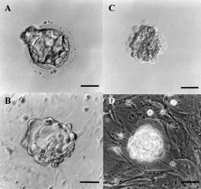
Fig. 1.
Immunosurgery of a human blastocyst for the derivation of human ES cell line. (A) Donated human embryo produced by in vitro fertilization at the blastocyst stage. (B) Human blastocyst after zona pellucida removal by Tyrode’s solution, during exposure to rabbit anti-human whole antiserum. (C) Embryo after exposure to guinea-pig complement. (D) Intact inner cell mass immediately after immunosurgery on mitotically inactivated mouse embryonic fibroblast feeder layer. Scale bar = 50 µm.

