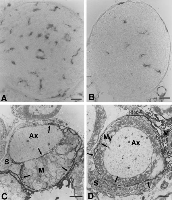
Fig. 2.
(A,B) Immunohistological localization of macrophages in femoral quadriceps nerves of P0+/− (A) and P0+/+ mice (B) at the age of 6 months using antibodies to F4/80. In quadriceps nerves of P0+/− mice the number of macrophages is clearly elevated when compared to P0+/+ mice. Note the larger size of the cells and the close vicinity of two cells to an endoneurial blood vessel in the nerve of the mutant (A). (C,D) Immunoelectron microscopic localization of F4/80-positive macrophages in peripheral nerves of 6-month-old P0+/− mice. (C) An F4/80-positive macrophage (M), containing myelin debris, is in close apposition to a demyelinated axon. Arrows indicate electron-dense immunoreaction product. Axon (Ax), Schwann cell (S). (D) A slender, immunoreactive process (arrows) of an F4/80-positive macrophage (M) has penetrated in between the pericaryon of a Schwann cell (S) and its normal appearing myelin sheath (My). Scale bars: 20 μm (for A and B); 1.5 μm (for C and D).

