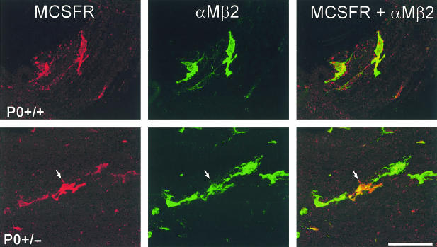
Fig. 4.
Cellular localization of the M-CSF receptor (MCSFR, red) immunoreactivity in teased fibre preparations from ventral roots of P0+/+ and P0+/− mice using antibodies to αMβ2 integrin (green) as a marker for peripheral nerve macrophages. αMβ2-negative cells, such as the adjacent Schwann cells, were never labelled. Note the particularly strongly labelled M-CSFR-immunoreactive macrophage in the P0+/− mutant (arrow). Scale bar: 50 μm.

