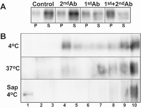
Fig. 1.

Antibody cross-linking leads to MAG partitioning into detergent-insoluble microdomains. (A) Western blot analyses of MAG from untreated OLs (control) or following incubation for 15 minutes with anti-mouse IgG (1:500, 2ndAb), anti-MAG IgG mAb (1:100, 1stAb) or anti-MAG mAb (1:100) followed by anti-mouse IgG (1:500, 15min; 1st+2ndAb). Cell lysate (10μg of protein) was extracted with TX-100 at 4°C, separated by centrifugation into insoluble (P) and soluble (S) fractions, and the entire yield in each fraction loaded on the gel. (B) OLs were extracted as above, with TX-100 at 4°C, at 37°C and at 4°C after pretreatment of the cell lysate with saponin. The insoluble fractions from MAG-cross-linked cells (anti-MAG + anti-mouse IgG, 50μg protein) were then further fractionated by centrifugation on sucrose gradients (see Methods). Fraction 1, is the top of gradient (i.e. lowest density). The figure represents a typical result of three independent experiments.
