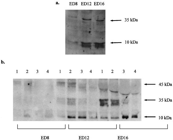
Fig. 1.
Western blots for caspase-9 immunoreactivity (antibody from Stressgen Inc.), showing fragments at 10 and 35 kDa indicative of activated caspase-9. Immunoreactivity is shown for lyates of whole lenses from ED-8, -12 and -16. Immunoreactivity for cleaved fragments is strong at ED-12 and ED-16, but weaker at ED-8. (b) Western blots for caspase-9 immunoreactivity (antibody from Stressgen Inc.), showing fragments at 10 and 35 kDa indicative of activated caspase-9. Immunoreactivity is shown for each of the four dissected regions of lenses at ED-8, -12 and -16. Expression of the 10-kDa fragment is seen primarily at ED-12 in regions 2 and 3, and at ED-16 in regions 3 and 4. Expression of pro-caspase-9, at 45 kDa, is seen at ED-12 and -16, but at ED-16 this band is displaced by the abundance of crystallin which is also expressed at this time.

