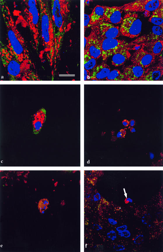
Fig. 5.
Lens epithelial cells cultured for 5 days to allow the differentiation of lentoids, and immunostained using the caspase-9 antibody from Stressgen Inc. Nuclei are labelled with DAPI (blue). Bar = 5 µm. (a) Undifferentiated cells from a lens cell culture stained with MitoTracker® (red) and anticaspase-9 (green). Caspase-9 is not localized to the mitochondria. (b) As a, but stained for cytochrome c (green) and caspase-9 (red). Cytochrome c and caspase-9 do not co-localize in non-differentiating cells. (c) A denucleating cell in a lentoid stained with MitoTracker® (red) and with an antibody to the 10-kDa fragment of caspase-9 (green). Activated caspase-9 is not present in normal mitochondria, which are in a perinuclear location. (d) As in c, stained for cytochrome oxidase (green) and caspase-9 (red), showing co-localization (yellow) in a perinuclear location. (e) As in c, stained for cytochrome c (green) and caspase-9 (red). The cytochrome c labelling is perinuclear but diffuse. (f) As in c and e, the denucleating cell (arrow) is stained for cytochrome c (green) and caspase-9 (red). Cytochrome c has disappeared, but caspase-9 persists.

