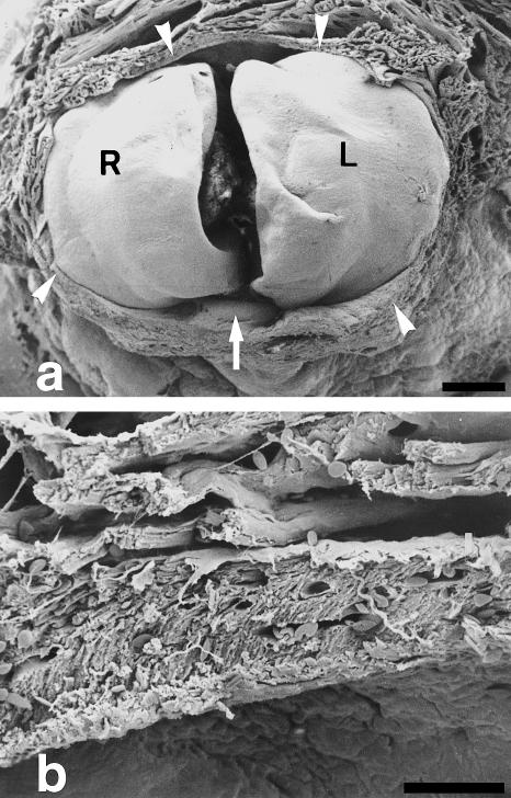
Fig. 2.
SEM micrographs depicting the junctional area between the ventricle and the bulbus arteriosus. (a) The ventricle has been dissected out and the caudal (ventricular) aspect of the left (L) and right (R) conus valves are exposed. Note the presence of a muscular ring (arrowheads) surrounding the valve leaflets. Arrow points to a posterior, supernumerary leaflet. (b) Detail of the anterior part of the muscular ring. It is formed of compact, vascularized myocardium. Myocytes are arranged in parallel and appear obliquely orientated. Vessels are more apparent on the outer side of the muscular ring. Myocardial trabeculae appear on the upper half of the micrograph. Scale bars = a, 200 μm; b, 30 μm.

