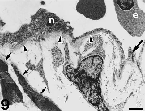
Fig. 9.
Detail of a myocardial vessel. Endothelial cells show dark nuclei (n), microvilli and a thick basement membrane (arrowheads). Tight junctions appear at the points of contact (large arrow). An interstitial cell appears in the subendocardium contacting abundant collagenous material. Myocytes show a well-developed basement membrane (small arrows). Erythrocytes (e) appear in the vessel lumen. Scale bar = 1 μm.

