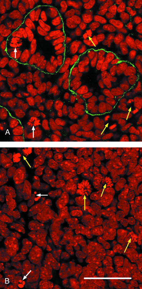
Fig. 2.
Optical sections through an E14.5 mouse kidney stained with propidium iodide (red) and anti-laminin antibody (green). (A) Nephric tubules embedded in stromal cells with two cells in metaphase (white arrows) and three cells in anaphase (yellow arrows). (B) Stem cells at the kidney periphery with two cells undergoing apoptosis (white arrows) and four cells in different stages of mitosis (yellow arrows). Scale bar = 40 μm.

