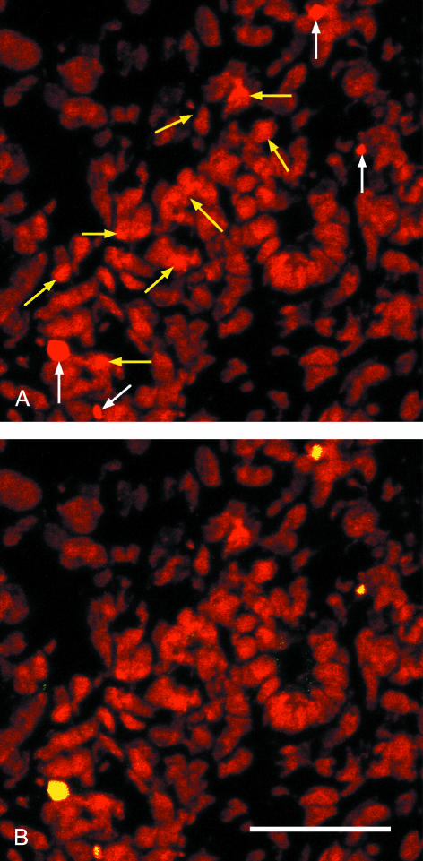
Fig. 4.
Confocal micrographs of a cryosectioned E14.5 kidney cortex stained with propidium iodide (red, A,B) and FITCTUNEL (yellow, B). (A) Propidium staining alone shows four intensely stained nuclei (white arrows) and several fragmented nuclei whose staining is a little more intense than the background (yellow arrows). (B) The FITC–TUNEL confirms that the four intense nuclei (A) are undergoing apoptosis but that none of the fragmented nuclei is apoptotic. Scale bar = 40 μm.

