
Supplementary Figure 1.
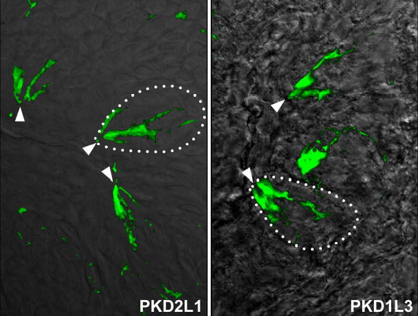
PKD2L1 and PKD1L3 are enriched in the taste pore
Immunofluorescent stainings of mouse taste buds with PKD2L1 (left panel) and with PKD1L3 (right panel) antibodies. The pictures show superposition of fluorescent antibody signals on DIC images of taste tissue. Dotted lines illustrate the outline of a taste bud, and arrows point to the taste pore region