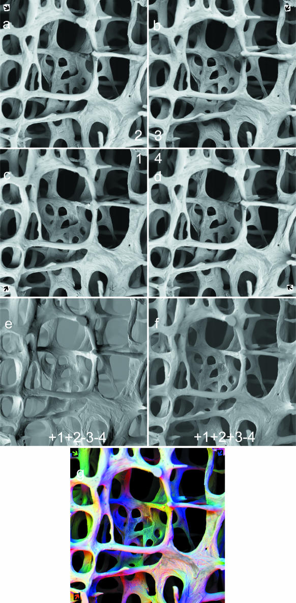
Fig. 1.
panels a–g: 90° sectors of annular solid-state BSE detector. 30 kV BSE scanning electron micrographs of 68-year-old human female L4 vertebral body trabecular bone (vertical slice cleaned of cells and coated with evaporated carbon to make it electrically conductive): secondary hyperparathyroidism of chronic renal failure accounts for patches of new, fine-trabecular bone and of superficial unmineralized ‘osteoid’ appearing darker in a–d and grey in g. Field width = 2.23 mm. Working distance from final lens (WD) = 25 mm: detector is 4.5 mm thick, 0.5 mm from lens. Thus sample to detector distance in this case = 19 mm. a–d: Top four panels: sector 1,2,3,4 BSE images of same field at same focus level in panels c,a,b,d, respectively, sector number in centre corner of cluster, arrow showing direction of apparent illumination due to that sector in outer corners. e: Lower left (1 + 2) – (3 + 4). f: Lower right + 1 + 2 + 3 – 4. panel g: R = 1↗, G = 2, B = 3 combination of panels c,a,b, respectively. Coloured arrows show direction of apparent illumination due to three 90° sectors.

