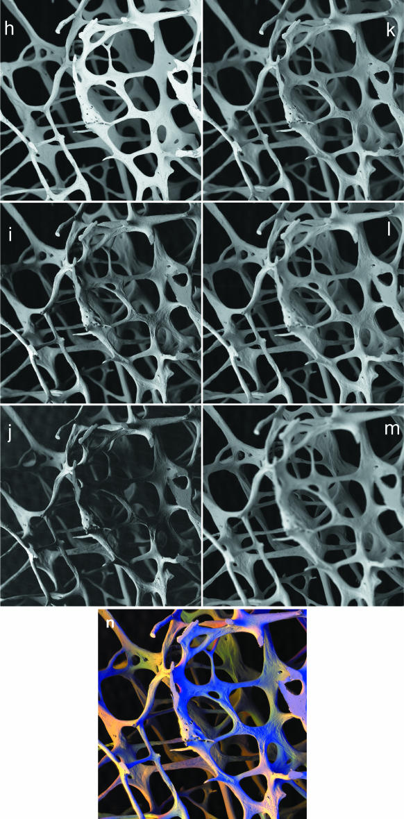
Fig. 2.
panels h–m: In, Off, Far positions employing all sectors of annular solid state BSE detector. 20 kV BSE scanning electron micrographs of 84-year-old human female L4 vertebral body trabecular bone. Twelve levels were recorded with the sample being moved mechanically 250 µm towards the detector at each step. At each level, the same detector was placed in three contrasting positions for three separate recordings. In position = (‘overhead’, annular detector surrounding the electron beam axis), Off-axis position, the first position at which the electron beam clears the BSE detector to one side. Far off-axis position = further movement of detector. Field width = 4.05 mm. WD = 23 mm. h,i,j: I,O,F images of same field at same focus level, 750 µm below top of sample. k,l,m: Combined IOF images at focus 250 µm, 1500 µm, 2500 µm below top of sample.panel n: After generating each colour composite image, the first 11 were changed to match the magnification to that of the last, and they were then processed using AutoMontage™ to extract an image in focus from back to front, where R = Far, G = Off, and B = In. Colour varies with surface slope and direction.

