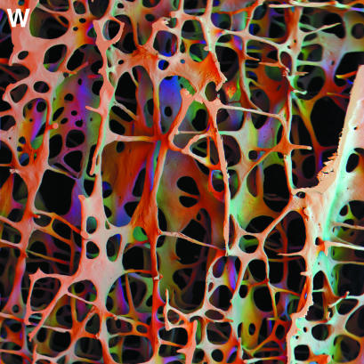
Fig. 4.
panel w: In, Off, Far positions employing all sectors of annular solid-state BSE detector. Scanning electron micrograph of 84-year-old male porotic bone. A 10-mm window in a 3-mm-thick vertical slice of fourth lumbar vertebral body trabecular bone, cleaned and coated with carbon: this is an exceptionally wide field of view (low magnification) image from an SEM. 30 kV. WD = 41 mm. In position (surrounding the electron beam axis) was assigned as red, a first off-axis position as green and a far off-axis position as blue in a combined image. Colour varies with surface slope and direction.

