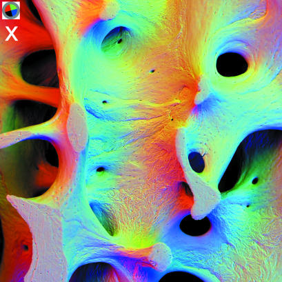
Fig. 5.
panel x: 90° sectors of annular solid state BSE detector. 20 kV BSE scanning electron micrographs of 31-year-old human male L4 vertebral body trabecular bone (vertical slice cleaned of cells and coated with evaporated carbon to make it electrically conductive). Field width = 2.70 mm, WD = 21 mm. Inset top left shows position of active detector sectors, i.e. direction of apparent illumination due to three 90° sectors. R = 2, G = 3, B = 4.

