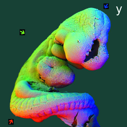
Fig. 6.
panel y: 90° sectors of annular solid-state BSE detector, to demonstrate that methods also work for conventionally prepared biological soft tissue SEM samples. 30 kV BSE scanning electron micrograph of 9-day post-fertilization mouse embryo, fixed in 3% glutaraldehyde in 0.2 m cacodylate buffer, post-fixed in 3% osmium tetroxide in same buffer, both for 16 h, dehydrated in ethanol, substituted with Freon-113 and critical point dried from carbon dioxide, mounted on spherical Aluminium rivet, sputter coated with gold. Fieldwidth = 1.85 mm. WD = 17 mm. Three of 4 angular sectors of an annular detector were used to obtain three different images which were then used to synthesize a colour image: coloured arrows show directions of apparent illumination. R = 1, G = 2, B = 3.

