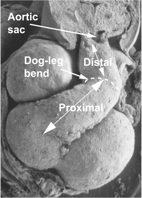
Fig. 4.
This scanning electron micrograph of a human embryo, at Carnegie stage 15 (approximately 34 days of gestation), shows a ventral view of the heart. The distal and proximal segments of the unseptated outflow tract are separated by a characteristic dog-leg bend. The proximal segment lies across the atrioventricular junction.

