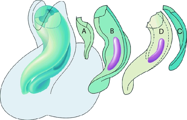
Fig. 10.
Reconstruction made from a human heart of 5 weeks gestation (Carnegie stage 15), viewed from the ventral aspect, soon after immigration of neural crest cells has begun. The left-hand panel shows the overall arrangement, with the arrangement of the individual cushions shown to the right. (A,C) Intercalated ridges or cushions, which occupy the area of the dog-leg bend; (B) septal outflow ridge; (D) parietal outflow ridge. Localized accumulations of neural crest-derived mesenchyme form ‘prongs’, shown in purple. Note that they are located within the distal parts of the cushions of the proximal outflow tract, being positioned just proximal to the dog-leg bend.

