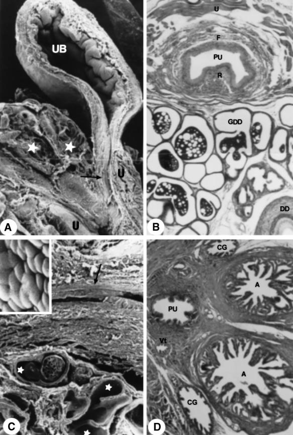
Fig. 2.
(A) Sagittal section through urinary bladder. Stars, glandular tissue; arrow, pelvic urethra. ×20. (B) Cross-section through pelvic urethra. MlT stain. ×455. (C) Glandular tissue detail (stars) and pelvic urethra (arrow). ×465. Inset: epithelium surface of the pelvic urethra. The superficial cells are irregularly polygonal. ×1110. (D) Cross-section through the pelvic urethra. MlT stain. ×202. For abbreviations, see text.

