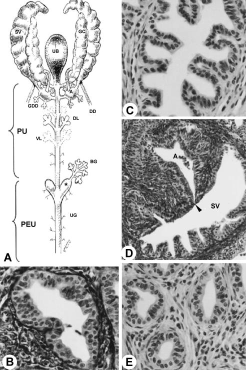
Fig. 4.
(A) Diagram of the ventral view of the sex accessory gland, ducts and urethra. Star, urethral diverticle. (B) Dorsal prostatic duct. MlT stain. ×1750. (C) Duct of the coagulating gland. HE stain. ×2285. (D) Opening (arrowhead) of the duct of the seminal vesicle (SV) into ampullary duct (A). MlT stain. ×895. (E) Ventral prostatic duct. HE stain. ×1644. For abbreviations, see text.

