
Fig. 13. Histochemistry and immunohistochemistry of the conus valves. B, bulbus; C, conus; S, sinus wall; SEp, subepicardial tissue.
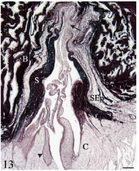
Orcein. Longitudinal section. The fibrous cylinder shows two different parts. The area corresponding to the sinus wall is very rich in elastin. The areas corresponding to the valvar attachments (arrow) are poor in elastin but rich in collagen. The subepicardial tissue also shows elastin fibres. Scale bar = 100 µm.