
Fig. 14. Histochemistry and immunohistochemistry of the conus valves. B, bulbus; C, conus; S, sinus wall; SEp, subepicardial tissue.
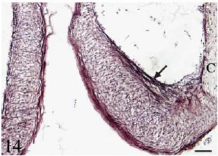
Orcein. Longitudinal section. Individual elastin fibres run longitudinally along the luminal side of the leaflets (arrow). Elastin fibres also course transversally from the parietal to the luminal side of the leaflet. Scale bar = 25 µm.