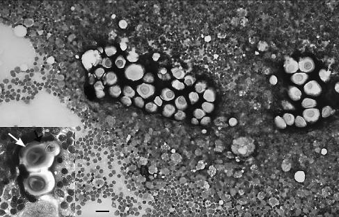
Figure 1.
Photomicrograph of a fine needle aspirate from the submandibular lymph node of a ferret with cryptococcosis. Cryptococcus neoformans vary in size, are surrounded by a negative staining capsule, and highlighted by a background population of lymphocytes and a few large macrophages. Wright-Giemsa stain; bar = 32 μm. Insert: Higher magnification of 2 Cryptococcus neoformans surrounded by lymphocytes. A bud with narrow base and surrounding capsule can be seen in the uppermost organism (arrow). Wright-Giemsa stain; bar = 14 μm.

