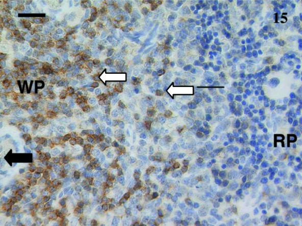
Fig. 15.
Spleen from a 38-day-old animal stained using anti-CD5. Obvious stained lymphocytes (white arrows) were seen in the white pulp (WP) areas compared with the red pulp (RP) area. Periarterial lymphatic sheaths (PALS) were evident in the white pulp as well as large cells (black arrow) in the red pulp. Scale bar, 25 µm.

