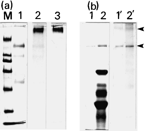
Fig. 3.
SDS–PAGE and immunoblotting of peri-albumen layer (PL) materials. (a) Isolated PL materials were separated through 7.5% SDS–PAGE and stained with Coomassie Brilliant Blue (CBB; lane 1), PAS (lane 2) or Alcian blue (lane 3). M, marker proteins of 200, 116, 97.4, 66, 45 and 31 kDa from top to bottom. (b) SDS–PAGE profiles of isolated PL materials (lanes 1 and 1′) and their crude preparations (lanes 2 and 2′), stained with CBB (lanes 1 and 2) or the antiserum (lanes 1′ and 2′) against the PL materials. The 260- and 160-kDa bands were specifically stained with the antiserum (arrowheads).

