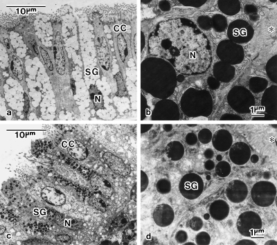
Fig. 7.
Ultrastructures of mucosa of the magnum (a,b) and isthmus (c,d). (a) Alternately distributed secretory cells and ciliated cells (CC) in luminal epithelium of the posterior portion of the magnum. The secretory granules (SG) are filled with fibrillar material of low electron density. (b) Secretory cells possessing electron-dense SG in tubular gland at the magnum. (c) Alternately distributed secretory cells and CC in luminal epithelium of the isthmus. SG is filled with materials of various electron density. (d) Secretory cells possessing electron-dense SG in tubular gland at the isthmus. Electron-lucent microstructures are seen in SG. The asterisk (*) indicates the glandular canal. N, nucleus.

