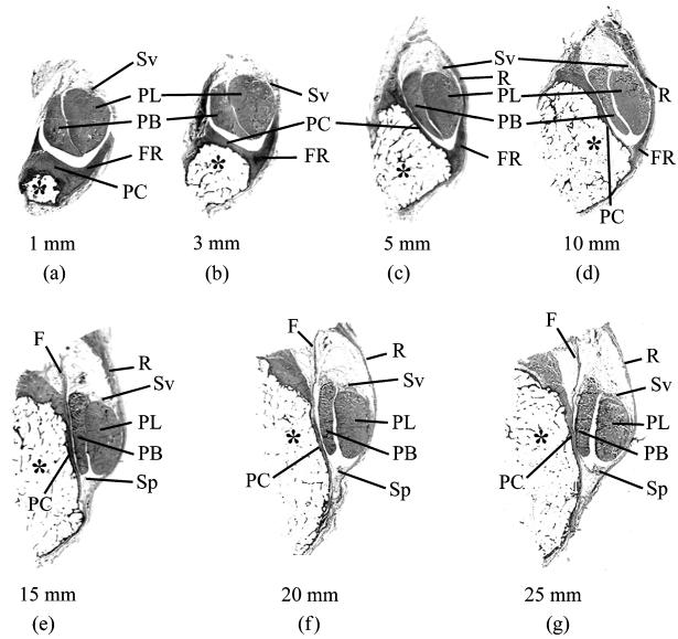
Fig. 1.
Macroscopic views of serial transverse sections through the malleolar groove and its contained tendons [peroneus brevis (PB) and peroneus longus (PL)]. The sections are taken at 1, 3, 5, 10, 15, 20 and 25 mm from the tip of the lateral malleolus (a–g, respectively). The top of each figure is posterior. At the distal end of the groove (1- and 3-mm sample points), the periosteal cushion (PC) is thick and together with the prominent fibrous ridge (FR) its shape compensates for the convexity of the lateral malleolus (*) in these regions. More proximally (i.e. at all other sample points), the groove is either flat or slightly concave, and the associated periosteal cushion is thus much less prominent. At all sample points 5 mm or more from the tip of the malleolus, the superior peroneal retinaculum (R) forms much of the lateral border of the groove. The synovial sheath associated with the peroneal tendons is present in all regions, but its parietal layer (Sp) is only visible at the 15- to 25-mm sample points. Although the visceral layer of the sheath (Sv) is present throughout, it is restricted to the sides of the tendons facing away from the bone. All sections are stained with Masson's trichrome. F is a band of fibrous tissue continuous with the purely fibrous periosteum at this proximal part of the malleolar groove.

