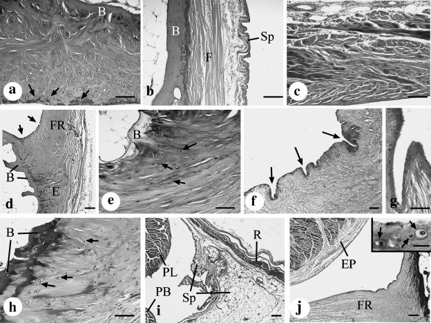
Fig. 2.
(a) The distal part of the malleolar groove (3 mm from its tip) is lined by a thick, fibrocartilaginous periosteal cushion that largely accounts for the concavity or flat appearance of the groove in this region. Note the presence of fibrocartilage cells throughout the cushion and especially near the ‘articular’ surface of the malleolar groove (arrows). B, bone. Haematoxylin & alcian blue. Scale bar, 100 µm. (b) The proximal part of the malleolar groove (20 mm from its tip) is lined by a thinner and purely fibrous periosteum (F) that lacks cartilage cells. Fused with the periosteum is the parietal layer of the synovial sheath (Sp) that is associated with the peroneal tendons. B, bone. Haematoxylin & alcian blue. Scale bar, 100 µm. (c) The purely fibrous character of the superior peroneal retinaculum demonstrated in a section taken 10 mm from the tip of the malleolus. Masson's trichrome. Scale bar, 100 µm. (d) The base of the fibrous ridge (FR) on the lateral aspect of the malleolar groove, in a section taken 5 mm from the malleolar tip. The prominent fibrocartilage at its enthesis (E) is shown at higher magnification in (e) and the microscopic folds (arrows) in (f) and (g). B, bone. Haematoxylin & alcian blue. Scale bar, 200 µm. (e) High-magnification view of the fibrocartilaginous enthesis at the attachment of the fibrous ridge to the bone (B) 5 mm from the tip of the malleolus. Note the prominent fibrocartilage cells (arrows). Toluidine blue. Scale bar, 100 µm. (f) Numerous microscopic folds (arrows) are present at the angle between the periosteal cushion and the base of the fibrous ridge in this section taken 1 mm from the tip of the malleolus. Toluidine blue, Scale bar, 100 µm. (g) High-magnification view of a microscopic fold that is present at the angle between the periosteal cushion and the base of the fibrous ridge in this section taken 3 mm from the tip of the malleolus. Haematoxylin & alcian blue. Scale bar, 50 µm. (h) Numerous fibrocartilage cells (arrows) at the enthesis of the posterior talofibular ligament, which forms the medial border of the malleolar groove in this section taken 3 mm from the tip of the malleolus. B, bone. Masson's trichrome. Scale bar, 100 µm. (i) The parietal layer of the synovial sheath (Sp) associated with peroneus longus (PL) and peroneus brevis (PB) is prominent on the inner aspect of the superior peroneal retinaculum (R) in this section taken 25 mm from the tip of the malleolus. Masson's trichrome. Scale bar, 200 µm. (j) By contrast to the proximal end of the groove (see i above), there is no parietal layer of the synovial sheath in its distal half (this section is 10 mm from the malleolar tip). Instead the sheath is replaced by the fibrous ridge (FR). The visceral layer of the synovial sheath is represented by the epitenon (EP) on the surface of the peroneal tendons. Masson's trichrome. Scale bar, 200 µm. Inset: conspicuous fibrocartilage cells (arrows) are present near the articular surface of the fibrous ridge, but there is no synovial membrane. Masson's trichrome. Scale bar, 20 µm.

