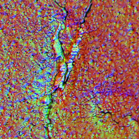
Fig. 3.
Medial condylar groove of 2-year-old Thoroughbred racehorse from MUGES training study, hyaline articular cartilage removed to the level of the mineralizing front (MF) of ACC using Tergazyme, carbon-coated, 20-kV SEM. BSE images recorded with three 90° sectors of annular solid-state BSE detector are used as RGB components (Boyde, 2003). Calcified material has filled a crack in ACC and is slightly proud of the MF. Field width, 900 µm.

