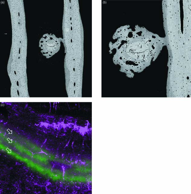
Fig. 13.
Fracture of transverse rod (centre) in parallel plate (each side of field) trabecular bone of 18-month-old Thoroughbred racehorse from the Bristol treadmill exercise training experiment, approximately 20 mm from distal articular surface. (a,b) 20-kV BSE SEM of central medio-lateral frontal section. (a) Field width, 1643 µm; (b) different micromilling level, old broken rod between four arrows, Field width, 530 µm. (c) Combined reflection (purple = red & blue) and fluorescence (calcein green, arrows) confocal image, showing 24-h increments of lamellar bone formed in initial repair process of this microfracture. Field width, 74 µm.

