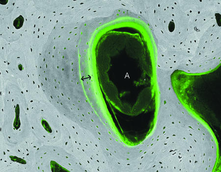
Fig. 15.
20-kV qBSE image of distal third metacarpal bone from Thoroughbred racehorse from the MUGES training study: image is combined with confocal fluorescence image to show calcein label groups, 3 weeks apart (double arrow), and histology in marrow space compartment: note the large arteriolar blood vessel (A). Field width, 1.3 mm.

