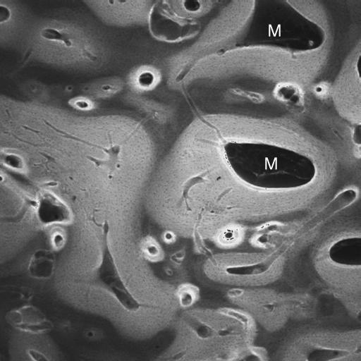
Fig. 16.
Same block as Fig. 13, low-magnification confocal laser scanning fluorescence image to show infilling of prior marrow space with new bone, which shows a brighter fluorescence level. M = residual marrow space. Field is 7 mm deep to medial condylar groove. Field width 2 mm.

