Fig. 4.
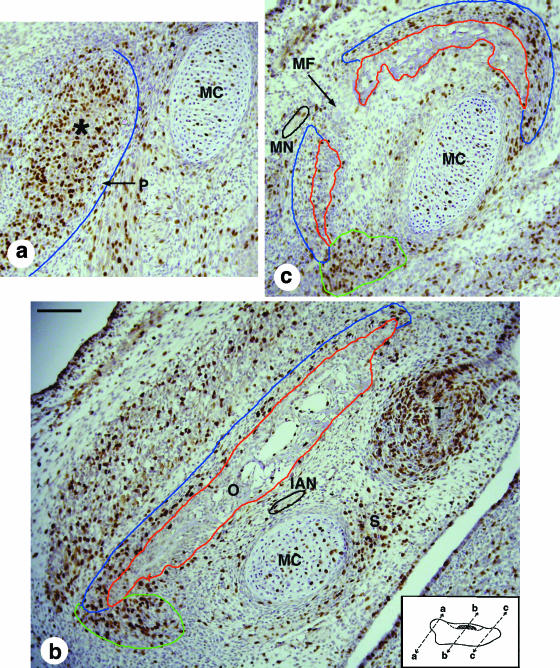
Micrographs of BrdU-stained sections of the E14.5 mandible, whose differentiated bone and periosteum are outlined in red and blue, respectively. Inset: the location and orientation of the sections, where proximal is to the left and superior is at the top. (a) Proximal region. The mesenchymal condensation that will give rise to the condylar cartilage (asterisk) forms within the developing perichondrium; although most of this is well defined, its lateral boundary is unclear and not outlined in blue. This condensation shows a high level of BrdU incorporation, in contrast to the low level seen in Meckel's cartilage. (b) Central region. The mandible is lateral to Meckel's cartilage, the inferior alveolar nerve (outlined in black) and the first molar tooth germ. BrdU incorporation is particularly strong in the inferior region of the mandible where there is one projection (outlined in green) towards Meckel's cartilage and another towards the tooth germ in the region of mesenchyme where the socket will form. The large BrdU-positive mesenchymal mass lateral to the mandible is the developing masseter muscle. (c) Distal region. This micrograph shows the mental foramen that separates the C-shaped mandible into two parts in this section. The mental nerve (outlined in black) is on the lateral side of the mandible in this micrograph. Note the high levels of BrdU staining in the mesenchyme at the inferior (outlined in green) and superior ends of the mandible where it extends towards Meckel's cartilage. IAN, inferior alveolar nerve; MC, Meckel's cartilage; MF, mental foramen; MN, mental nerve; O, osteoid tissue; P, periosteum; S, tooth socket; T, molar tooth germ. Scale bars, 100 µm.

