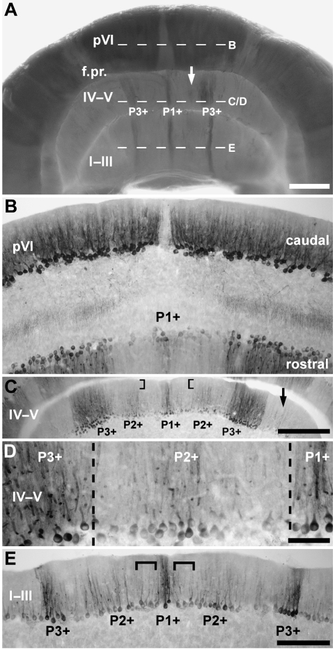
Fig. 4.
The topography of the anterior zone (AZ) of the vermis. (A) Zebrin II-immunoreactive Purkinje cells in the AZ, shown in whole mount. An array of zebrin II parasagittal stripes extends through all lobules anterior to the primary fissure (f.pr.). The arrow indicates the weak P2+ stripe and dashed lines indicate the approximate levels of the sections shown in B–E. (B) Horizontal section through the boundary between the AZ and CZ in lobule pVI, immunoperoxidase-stained for zebrin II. A narrow stripe of zebrin II-reactive Purkinje cells (P1+) straddles the midline on the rostral aspect. (C,D) Horizontal sections through lobule IV–V immunoperoxidase-stained for zebrin II. (C) Seen at lower magnification, the most striking stripe in the AZ is the thick (∼375 µm) heavily reactive P3+. Note that almost no zebrin II immunoreactivity is detected in the distal dendrites of Purkinje cells located within P2+ (square brackets), perhaps explaining why P2+ is not seen in the whole mount view (A). Patches of zebrin II-immunoreactive Purkinje cells lateral to P3+ are inconsistently stained and may represent P4+ (arrow). (D) Seen at high magnification, P1+ at the midline is flanked by P2+ and P3+ laterally. (E) Horizontal section through lobule I–III immunoperoxidase-stained for zebrin II. P1+ and P3+ are both strongly zebrin II immunoreactive and equal in width. P2+ is more weakly immunoreactive. At this rostrocaudal level, P1− is difficult to discern as the lateral border of P1+ and the medial border of P2+ merge (square brackets). Scale bars: A = 1 mm; C = 500 µm; D = 100 µm; E = 250 µm (also applies to B).

