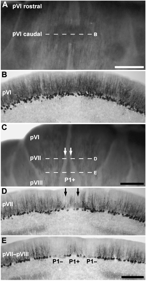
Fig. 5.
The topography of the central zone (CZ) of the vermis. (A) Dorsal view of the rostral half of lobule VI–VIII, presumptive lobules VI and VII (pVI–pVII), shown in an anti-zebrin II-stained whole mount preparation. Zebrin II is expressed throughout the CZ. (B) Horizontal section through pVI–pVII, immunoperoxidase-stained for zebrin II (the corresponding region is indicated by dashed line B in panel A). All Purkinje cells express zebrin II. (C) Dorsoposterior view of lobule VI–VIII, shown in whole mount. Between lobules pVI and pVII, P1+/P1− stripes, which appears to derive from the caudal PZ, interrupts the homogeneous zebrin II expression domain. (D) Horizontal section through lobule pVII, immunoperoxidase-stained for zebrin II (indicated by dashed line D in panel C). Peroxidase reaction product is heavily deposited in most Purkinje cell dendrites, somata and axons but dendritic immunoreactivity is weak in a pair of stripes adjacent to the midline (arrows). (E) Horizontal section through lobules pVII–pVIII immunoperoxidase-stained for zebrin II (indicated by the dashed line E in panel C). P1+ becomes clearly differentiated from the immunopositive Purkinje cells located laterally. Scale bar in A = 1 mm; C = 1 mm; E = 250 µm (also applies to B and D).

