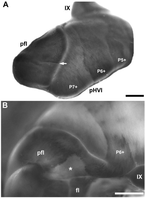
Fig. 9.
Zebrin II expression in the paraflocculus and flocculus shown in whole mount immunoperoxidase-stained cerebella. (A) Lateral view of the paraflocculus (pfl). The heavily reactive Purkinje cells in the posterior paraflocculus form a sharp boundary (arrow) with the weakly reactive anterior paraflocculus. pHVI and vermian lobule IX are labelled. (B) Ventroposterior view of the paraflocculus and flocculus (fl). All Purkinje cells express zebrin II. The P6+ stripe extending over the hemispheres of the posterior hemispheric lobules appears to be continuous with the paraflocculus. A large cell-poor area in the posterior paraflocculus (asterisk) presents as an immunonegative gap in the zebrin II expression. Ventrally, lobules IX and X (hidden from view) of the vermis are morphologically continuous with the paraflocculus and the flocculus, respectively. Scale bars = 1 mm.

