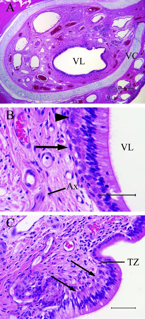
Fig. 1.
The VNO (A) is associated with numerous blood vessels and glands that are ensheathed by the vomeronasal cartilage (scale bar = 300 µm). The SE (B) contains sensory neurones (arrowhead) in a layer 1–3 cell bodies thick. The neurones are immediately apical to basal cells (arrow) that lie on the basement membrane. Axons fasciculate (Ax) in the lamina propria (scale bar = 50 µm). In (C), the sensory epithelium (arrows) abuts the non-sensory epithelium at the transitional zone (scale bar = 50 µm).

