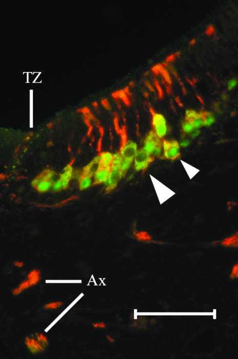
Fig. 4.
A layer of 1–3 PGP(+) cells occupies the basal compartment of the sensory epithelium. A section double-labelled with BT in red (Alexa 594) and PGP in green (Alexa 488) reveals that the sensory neurones do not label consistently with each antibody. Cell somata that are clearly BT(+)/PGP(+) are yellow in colour (small arrowhead). Some dendrites and axon bundles contain both labels. Many nuclei are PGP(+) (small arrowhead) but some nuclei are PGP(−). At the TZ, small structures in the NSE are BT(+) and/or PGP(+) (scale bar = 50 µm).

